October 2005

Mass around the aorta

Clinical information

Impaired renal function

Ultrasound Images & Clips

70 years old male, Impaired renal function

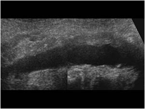
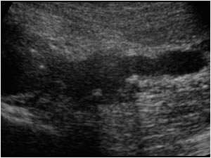
Longitudinal image showing a mass ventral of the distal abdominal aorta and a normal proximal aorta.
Transverse image showing the mass around the distal aorta.
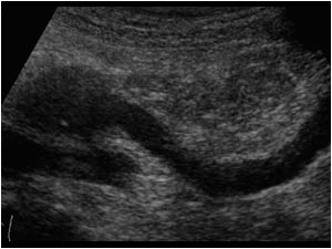
Longitudinal image of the right kidney with a dilatated collecting system.
Transverse image of the right kidney with a dilatated collecting system.
Longitudinal image of the left kidney with a dilatated collecting system.
Transverse image of the left kidney with a perirenal effusion caused by a fornix rupture.
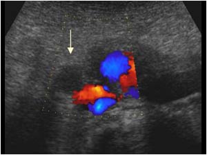
Transverse color doppler image showing a medial displaced and dilatated right ureter ventral of the vena cava and next to the aortic mass (arrow).
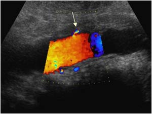
Longitudinal color doppler image of the aorta with ventral mass and origin of the inferior mesenteric artery (arrow).
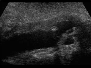
Longitudinal image of the distal aorta and proximal left common iliac artery showing the mass extending along the proximal left common iliac artery.
Longitudinal image of the left common iliac artery showing the mass extending along the proximal left common iliac artery. The distal common iliac artery is normal.
Longitudinal image of the right common iliac artery showing the mass extending along the proximal right common iliac artery. The distal common iliac artery and external iliac artery are normal.
CT image at the level of the kidneys showing the right kidney with a dilatated collecting system and the left kidney with a perirenal effusion. The aorta at this level is normal.
CT image showing the mass around the aorta.
CT image showing the mass around the proximal right and left common iliac artery.
Plain abdominal X ray showing a stent in the right and left ureter.

The above images present the typical characteristics off a case of retroperitoneal fibrosis. Retroperitoneal fibrosis often presents with a dilatation of the renal collecting system ca
Plain abdominal X ray showing a stent in the right and left ureter. The above images present the typical characteristics off a case of retroperitoneal fibrosis. Retroperitoneal fibrosis often presents with a dilatation of the renal collecting system ca

Details

  • Sex: Male
  • Age: 70

Created with

  • Fujifilm Ultrasound System