November 2005

Atypical cyst in the breast

Clinical information

Palpable mass with benign characteristics

Ultrasound Images & Clips

68 years old female, Palpable mass with benign characteristics

Longitudinal image of a thick walled cyst with slightly irregular contour (left side of the image).
Transverse image of the same cyst.
Mammographic image of the cyst. Note the irregular contour of the lower part of the lesion.
Cranio-caudal view of the same lesion.
There is no hypervascularity of the cyst wall.
An ultrasound guided puncture of the cyst was performed. The fluid that was aspirated was hemorrhagic. The patient was relieved because the lump had disappeared.
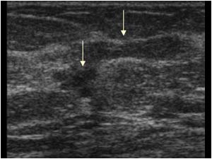
Careful examination after the cyst puncture however revealed a 5 mm small stellate lesion (lower left arrow) next to the empty cyst (upper right arrow).

The cytological examination of the cyst fluid did not show malignant cells. Because of the stellate
Careful examination after the cyst puncture however revealed a 5 mm small stellate lesion (lower left arrow) next to the empty cyst (upper right arrow). The cytological examination of the cyst fluid did not show malignant cells. Because of the stellate
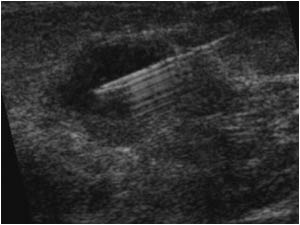
Longitudinal image of the cyst and the stellate lesion after two weeks. The cyst is already partly filled with fluid. The fluid in the cyst was again aspirated and a biopsy was performed of the thick wall of the cyst.

Also of course a biopsy was perfor
Longitudinal image of the cyst and the stellate lesion after two weeks. The cyst is already partly filled with fluid. The fluid in the cyst was again aspirated and a biopsy was performed of the thick wall of the cyst. Also of course a biopsy was perfor
5 mm stellate lesion and biopsy needle in pre-fire position.
5 mm stellate lesion and biopsy needle through the lesion.

The biopsy results of the cyst wall revealed no signs of malignancy. The biopsy of the stellate lesion however proved this mass to be malignant.
In cases of an atypical irregular thick walled
5 mm stellate lesion and biopsy needle through the lesion. The biopsy results of the cyst wall revealed no signs of malignancy. The biopsy of the stellate lesion however proved this mass to be malignant. In cases of an atypical irregular thick walled

Details

  • Sex: Female
  • Age: 68

Created with

  • Fujifilm Ultrasound System