January 2006

Hypoechoic lesion in the knee

Clinical information

Peroneal nerve palsy. The neurologist found a palpable lesion on the dorso lateral aspect of the knee.
Ultrasound Images & Clips

Male, 42, truck driver, Peroneal nerve palsy. The neurologist found a palpable lesion on the dorso lateral aspect of the knee.

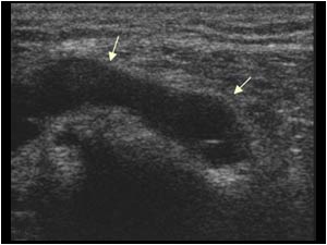
Longitudinal image of the dorso lateral aspect of the knee shows a tubular fluid filled structure.
Longitudinal image of the distal end of the lesion. The structure has a blind end and shows no flow .A vascular structure was ruled out.
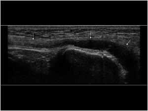
Longitudinal image of the same lesion. The structure is in relation with the peroneal nerve (proximal arrow).
Longitudinal image showing a thickening of the peroneal nerve in the distal direction.
Transverse image of the cystic lesion shows an oval lesion in close contact to the fibular head.
Three transverse images of the peroneal nerve from proximal to distal showing the close relation between the cystic lesion and the peroneal nerve.
There is not only a close relation between the cystic lesion and the peroneal nerve, but also to the tibiofibular joint space (proximal arrow).

Conclusion

It is well known that ganglia arising from the tibiof ibular joint space can cause peroneal nerve compression. In this case there seemed to be not only compression of the nerve but an actual relation of the lesion to the nerve. The patient was operated in a university hospital. The lesion proved to be a ganglion cyst of the peroneal nerve sheath. After removal of the lesion the peroneal nerve palsy resolved quickly.

For more peripheral nerve lesions see cases 7.3.3, for more tibiofibular ganglia see cases 6.6.8:

Details

  • Sex: Male
  • Age: 42

Created with

  • Fujifilm Ultrasound System