July 2007

Abnormal testis and epididymis

Clinical information

Patient is treated for an epididymitis for some time without result. There is persistent scrotal swelling and pain.
Ultrasound Images & Clips

22 year old African male, Patient is treated for an epididymitis for some time without result. There is persistent scrotal swelling and pain.
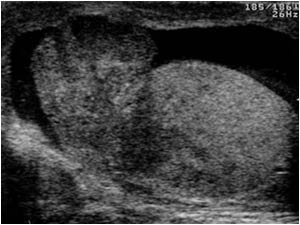
Longitudinal image of the left scrotum with an enlarged inhomogeneous epididymis and testicle, reactive hydrocele
Longitudinal image of the right scrotum with a normal testis
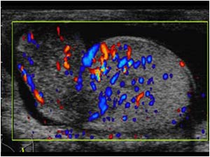
Longitudinal image of the left scrotum with an enlarged inhomogeneous hypervascularized epididymis and testicle.
Longitudinal image of the left testis with an enlarged inhomogeneous hypervascularized testicle.
There is even a hypervascularized appendix testis
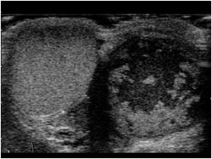
Transverse image of the right scrotum with a normal testis, but with an irregular peritesticular fluid filled mass representing caseous necrosis
Transverse image of the right scrotum with a normal vascularized testis. No flow in the irregular peritesticular fluid filled mass.
Transverse image of the lower abdomen with swollen hypoechoic seminal vesicles

Conclusion

The epididymo-orchitis on the left side and an inhomogeneous fluid filled epididymal mass on the right side caused by caseous necrosis and the swollen seminal vesicles all with poor response to conventional treatment were suspicious for tuberculosis. Patient had no signs of tuberculosis elsewhere in the body.

A sperm analysis however confirmed the diagnosis Genital tuberculosis.

References     
de Cassio Saito O, de Barros N, Chammas MC, Oliveira IR, Cerri GG.Ultrasound of tropical and infectious diseases that affect the scrotum.
Ultrasound Q. 2004 Mar;20(1):12-8.

Türkvatan A, Kelahmet E, Yazgan C, Olçer T. Sonographic findings in tuberculous epididymo-orchitis.
J Clin Ultrasound. 2004 Jul-Aug;32(6):302-5.

Details

  • Sex: Male
  • Age: 22

Created with

  • Fujifilm Ultrasound System