August 2007

Abnormal portal confluence and splenic vein

Clinical information

Vomiting and weight loss
Ultrasound Images & Clips

Female 76 years old, Vomiting and weight loss
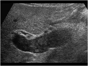
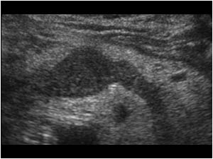
Transverse image of an abnormal portal confluence and splenic vein filled with a hypo-echoic intraluminal mass.
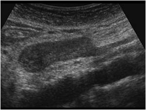
Longitidinal image of an abnormal superior mesenteric vein anterior of the aorta filled with a hypo-echoic intraluminal mass.
Transverse image of the portal vein with the same hypoechoic slightly lobulated hypo-echoic intraluminal mass.
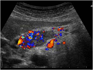
Transverse color doppler image of the portal confluence demonstrating flow in the intraluminal mass.
Cineloop shows flow in the intraluminal mass with e-flow
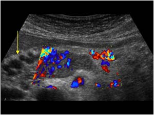
Transverse color doppler image of the portal confluence demonstrating flow in the intraluminal mass. There are tortuous cystic like structures visable next to the portal confluence.
Transverse color doppler image shows flow in the tortuous cystic like structures next to the portal confluence. They represent collateral veins. No primary tumor could be demonstrated with ultrasound.
CT scan demonstrating the mass in the portal vein and portal confluence.
CT scan demonstrating tumor in the splenic confluence and flow in the collateral veins.
Contrast study of the duodenum showing an obstructive mass in the inferior horizontal part of the duodenum.

Conclusion

Surgery proved an obstructing duodenal carcinoma with intravenous tumor extension. Because the tumor was irresectable a gastroenterostomy was performed to allow passage of food.

An intraluminal mass in the mesenteric or portal veins can be caused by a venous thrombosis or a tumor thrombus. Color doppler can help in the diagnosis by confirming or excluding flow in the mass.        

References   
Ishida H, Konno K, Hamashima Y, Naganuma H, Komatsuda T, Sato M, Kimura H, Ishida J, Sakai T, Watanabe S. Portal tumor thrombus due to gastrointestinal cancer.
Abdom Imaging. 1999 Nov-Dec;24(6):585-90. Review.

Details

  • Sex: Female
  • Age: 76

Created with

  • Fujifilm Ultrasound System