February 2008

Abnormal aspect of the median nerve

Clinical information

Palpable mass on the volar aspect of the hand and signs of median nerve compression
Ultrasound Images & Clips

22 year old female, Palpable mass on the volar aspect of the hand and signs of median nerve compression
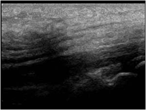
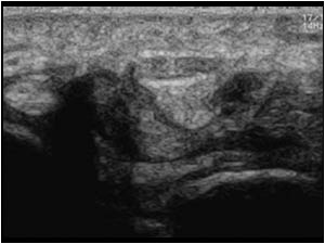
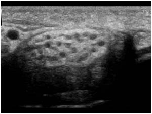
Transverse image of the median nerve proximal to the carpal tunnel. The nerve is massively thickened with normal and slightly thickened hypoechoic fascicles interspersed with hyperechoic material.
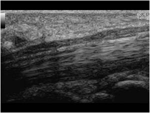
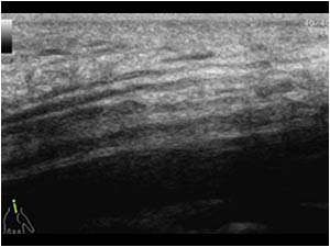
Longitudinal image of the median nerve proximal to the carpal tunnel showing the massively thickened nerve with normal and slightly thickened hypoechoic fascicles interspersed with hyperechoic material.
Longitudinal image of the median nerve in the carpal tunnel showing compression and calibre change of the massively thickened nerve.
Longitudinal image of the median nerve in the distal lower arm. The aspect returns to normal.
Transverse image of the median nerve in the distal lower arm. The aspect returns to normal.
Transverse image of the median nerve on the contra lateral side. The aspect of the median nerve is normal.
Longitudinal image of the median nerve on the contra lateral side. The aspect of the median nerve is normal.
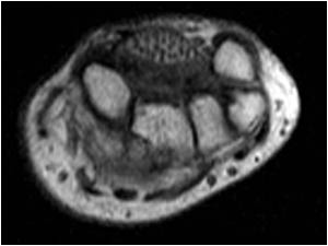
Transverse MRI image of the median nerve also showing the massively thickened nerve with normal fascicles interspersed with fatty material.
Longitudinal MRI image of the median nerve also showing the massively thickened nerve with normal and thickened fascicles interspersed with fatty material and carpal tunnel compression.
Longitudinal image of the thickened nerve made during operation.
Longitudinal image of the thickened nerve with slightly thickened fascicles interspersed with fatty tissue made during operation.

Conclusion

A carpal tunnel release was performed. Both the surgical and pathological results only demonstrate the presence of fatty tissue in the epineurium.
The condition is known as neural lipofibroma, neural lipoma or fibrolipomatous hamartoma.
It is a condition that mainly affects the median nerve. Usually the patients are young and sometimes the condition is associated with local gigantism (not the case here).

References            
Bianchi S,  Martinoli C. Ultrasound of the musculoskeletal system  page 101 and 489

Nouira K, Belhiba H, Baccar S, Miaaoui A, Ben Messaoud M, Turki I, Cheour I, Menif E. Fibrolipoma of the median nerve.
Joint Bone Spine. 2007 Jan;74(1):98-9.

Details

  • Sex: Female
  • Age: 22

Created with

  • Fujifilm Ultrasound System