August 2009

Abnormal right kidney

Clinical information

Urinary tract infection
Ultrasound Images & Clips

2 year old female, Urinary tract infection
Longitudinal image of the right kidney with a severely dilatated upperpole moiety
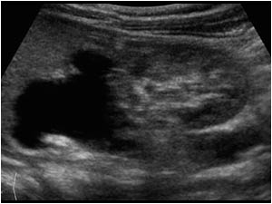
Longitudinal image of the left kidney without dilatation, but with a parenchymal bridge dividing the central complex indicating a duplex collecting system
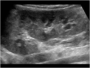
Longitudinal image of the right kidney with severely dilatated upperpole calices, renal pelvis and ureter. There is a normal amount of renal parenchyma in the lowerpole, but severe parenchymal loss in the upperpole.
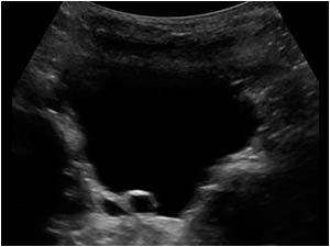
Longitudinal image of the bladder with a dilatated ureter of right upperpole
Transverse image of the bladder with a dilatated ureter of right upperpole ending in a small uretrocele.
Transverse high resolution image of the upperpole of the right kidney showing severe parenchymal loss in the upperpole
Transverse high resolution image of the lowerpole of the right kidney showing a normal parenchymal thickness

Conclusion

The diagnosis is a duplex collecting system on both sides, but with a dilatated upperpole moiety with parenchymal loss on the right side ending in an ureterocele.
The Weigert-Meyer Rule
Complete ureteral duplication with each segment having its own ureteral orifice in the bladder.
Upper Pole Moiety
Inserts medial and inferiorly to the lower pole moiety
Frequently ends in a ureterocele
Lower Pole Moiety
Inserts lateral and superiorly to the ureter draining the upper pole
Reflux can occur. (but in this case no reflux was found)
Usually, upper pole nephroureterectomy is performed for a nonfunctioning moiety (as in this case). For functioning segments, ureteropyelostomy or ureteric reimplantation can be considered.        

See also category 9.2.1 pediatric urinary tract with a case of complete ureteral duplication with an upperpole ending in an ureterocele and with reflux in the lowerpole system.            

Details

  • Sex: Female
  • Age: 2

Created with

  • Fujifilm Ultrasound System