October 2009

Abnormal gallbladder

Clinical information

An ultrasound examination was requested to exclude gallstones in a patient with vague abdominal pain
Ultrasound Images & Clips

32 year old female, An ultrasound examination was requested to exclude gallstones in a patient with vague abdominal pain
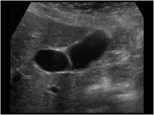
Longitudinal image of the gallbladder with thin septations across the gallbladder.
Longitudinal image of the gallbladder at another level showing the thin septations across the gallbladder.
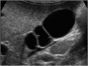
Transverse image of the same gallbladder with the septations forming a honeycomb appearance.
Transverse image of the same gallbladder at another level.
No stones could be found. All the septations are thin. There is no gallbladder wall thickening and there are no signs of a cholecystitis.
In this case the diagnosis is a multiseptate gallbladder
Transverse image of the same gallbladder at another level. No stones could be found. All the septations are thin. There is no gallbladder wall thickening and there are no signs of a cholecystitis. In this case the diagnosis is a multiseptate gallbladder
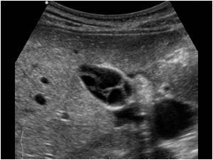
This is a longitudinal image of the gallbladder of a 54 year old female with only one septum within the gallbladder.
Image of the septum in another plane. The septum must be differentiated from a fold in the gallbladder which is a very common finding.
Another abnormally that can cause intraluminal septated structures is the membranous cholecystitis.
Image of the septum in another plane. The septum must be differentiated from a fold in the gallbladder which is a very common finding. Another abnormally that can cause intraluminal septated structures is the membranous cholecystitis.
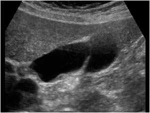
This is a longitudinal image of a membranous cholecystitis.
The intraluminal structures are caused by the sloughed mucosal lining of the gallbladder. The structures are more irregular and thicker than the septations of a multi septated gallbladder.
This is a longitudinal image of a membranous cholecystitis. The intraluminal structures are caused by the sloughed mucosal lining of the gallbladder. The structures are more irregular and thicker than the septations of a multi septated gallbladder.
In this case there is not only thickening of the gallbladder wall, but there are also clinical signs of a cholecystitis.

Conclusion

References            
Rivera-Troche EY, Hartwig MG, Vaslef SN. Multiseptate gallbladder. J Gastrointest Surg. 2009 Sep;13(9):1741-3.
Nakazawa T, Ohara H, Sano H, Kobayashi S, Nomura T, Joh T, Itoh M. Multiseptate gallbladder: diagnostic value of MR cholangiography and ultrasonography. Abdom Imaging. 2004 Nov-Dec;29(6):691-3.
Hahm KB, Yim DS, Kang JK, Park IS. Cholangiographic appearance of multiseptate gallbladder: case report and a review of the literature. J Gastroenterol. 1994 Oct;29(5):665-8.
Lev-Toaff AS, Friedman AC, Rindsberg SN, Caroline DF, Maurer AH, Radecki PD. Multiseptate gallbladder: incidental diagnosis on sonography. AJR Am J Roentgenol. 1987 Jun;148(6):1119-20.

Details

  • Sex: Female
  • Age: 32

Created with

  • Fujifilm Ultrasound System