March 2010

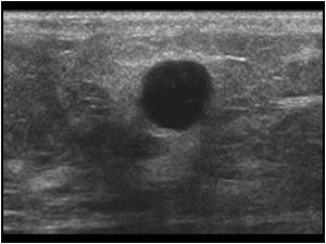
Intracystic mass in the breast

Clinical information

Palpable mass in the breast
Ultrasound Images & Clips

31 year old female, Palpable mass in the breast
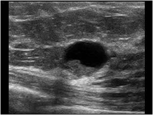
Intracystic lobulated mass in an ill defined cyst with an irregular wall
The same lesion in a different plane
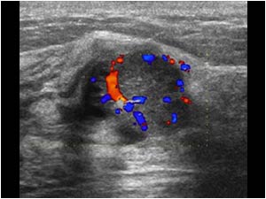
The mass is vascularized indicating that it is a tumor and not sludge
Both the tumor and cyst wall are vascularized.
Biopsy proved the lesion to be an intracystic ductal carcinoma.
Both the tumor and cyst wall are vascularized. Biopsy proved the lesion to be an intracystic ductal carcinoma.
Sometimes the lesion looks less suspicious as in this case of a complex cyst in the breast of a 73 year old female patient. There is a thin walled cyst with a mural nodule that did not move when the patient changed position. Although no vascularity was fo
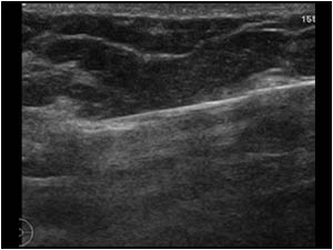
This image shows the vacuum biopsy needle direct underneath the abnormal cyst
After the procedure the cyst was completely removed and a marker was left in place.
The biopsy specimen showed an intracystic papilloma with ductal carcinoma in situ.
After the procedure the cyst was completely removed and a marker was left in place. The biopsy specimen showed an intracystic papilloma with ductal carcinoma in situ.
When dealing with breast cysts, optimal settings are very important. This less than 1 cm. breast lesion was found in a 67 year old female patient and initially interpreted as a simple benign cyst.
With different settings however the same lesion looks inhomogeneous and partly solid
The color Doppler proves the lesion to be vascularized
In the axilla of the same patient also an abnormal lymph node were found.
A biopsy procedure of the mass and the lymph node showed an infiltrating ductal carcinoma.
In the axilla of the same patient also an abnormal lymph node were found. A biopsy procedure of the mass and the lymph node showed an infiltrating ductal carcinoma.
This image shows two small cysts, one simple cyst and one atypical cyst. Fortunately not all atypical cyst are malignant. In this case it was a benign infected cyst.

Conclusion

When examining the breast with ultrasound, cysts are a very common finding. Be careful with atypical or complex cysts. Use optimal settings and use color Doppler to detect flow. When there is any doubt, perform a pathological examination.

Details

  • Sex: Female
  • Age: 31

Created with

  • Fujifilm Ultrasound System