April 2010

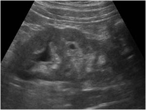
Abnormal structure in the fundus of the bladder.

Clinical information

Chronic urinary tract infection and hematuria
Ultrasound Images & Clips

Male patient 54 year old, Chronic urinary tract infection and hematuria
Longitudinal image of the bladder with an unusual structure in the fundus of the bladder.
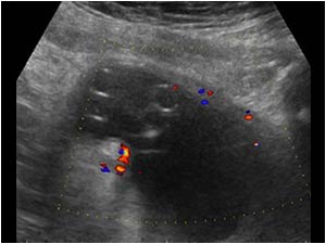
Transverse image of the same structure in the bladder.
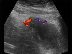
There is no vascularity within the structure.
The structure does not look like a polyp or transitional carcinoma.
Because the structure looked a bit like a part of a catheter, we asked the patient about a previous catheterization of the bladder. He deni
There is no vascularity within the structure. The structure does not look like a polyp or transitional carcinoma. Because the structure looked a bit like a part of a catheter, we asked the patient about a previous catheterization of the bladder. He deni
There is an increased vascularity in the bladder wall indicating a cystitis.
Because the kidneys are always included in the ultrasound examination of the urinary tract we also examined the kidneys.
There is an increased vascularity in the bladder wall indicating a cystitis. Because the kidneys are always included in the ultrasound examination of the urinary tract we also examined the kidneys.
Longitudinal image of the right kidney with a hypoechoic mass in the lowerpole of the kidney.
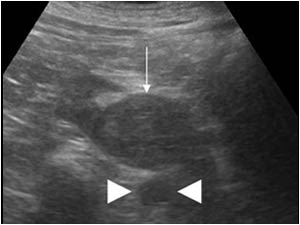
Transverse image of the mass in the lowerpole of the right kidney (thin arrow).
Also notice the position of the mass in relation to the aorta (arrow points).
Transverse image of the mass in the lowerpole of the right kidney (thin arrow). Also notice the position of the mass in relation to the aorta (arrow points).
Longitudinal image of the left kidney. The lowerpole is not well defined. Can you think of an abnormality that can explain this fact and the position of the tumor anterior of the aorta?
Of course a CT scan was performed.
Longitudinal image of the left kidney. The lowerpole is not well defined. Can you think of an abnormality that can explain this fact and the position of the tumor anterior of the aorta? Of course a CT scan was performed.
CT scan of the lowerpole of the kidneys. There is a horseshoe kidney. The mass in the lowerpole of the right kidney is clearly seen and is located anterior of the aorta.
Coronal CT image in the late phase, showing again the horseshoe kidney and the tumor.
In the lower abdomen the abnormal bladder can be seen. The intraluminal structure has a low density on CT.

Conclusion

During cystoscopy a foreign body in the bladder was found. The patient admitted auto manipulation during which a silicone string was lost in the bladder. The foreign body could be removed endoscopically. The renal mass proved to be a renal cell carcinoma, that had been incidentally discovered. The normal treatment for a renal cell carcinoma is a nephrectomy. Because the patient in this case has a horseshoe kidney, a nephrectomy is not possible. The patient was referred to a specialized urological center for a partial nephrectomy.

Details

  • Sex: Male
  • Age: 54

Created with

  • Fujifilm Ultrasound System