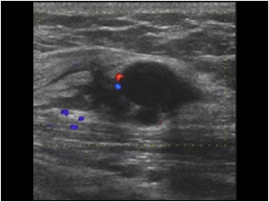
Painful palpable mass

Although both lesions look different on ultrasound, they are both an endometriosis in the abdominal wall. The clinical presentation of an endometriosis on the abdominal wall can vary. Many lesions are palpable, but the clinical presentation can be only pain.
In the classical case of an endometriosis the patient has localized pain during menstruation, but this is not always the case and pain can also be non cyclic.
The ultrasound presentation can vary . Some lesions are mainly solid and some are partly cystic. Vascularity is usually, but not always present.
When the lesion is mainly cystic, the lesion must be differentiated from a hematoma or seroma. When the lesion is ill defined it can mimic a malignant lesion.
Another common mass lesion in the abdominal wall is a desmoid tumor.
The presence of a mass lesion in the periphery of a scar after a cesarean delivery however makes the diagnosis endometriosis most likely. The time between the cesarean delivery and the clinical presentation is usually a few years. Some cases of endometriosis without previous surgery have been described.
An ultrasound guided biopsy can confirm the diagnosis. The preferred treatment is surgical removal.
References
Hensen JH, Van Breda Vriesman AC, Puylaert JB.
Abdominal wall endometriosis: clinical presentation and imaging features with emphasis on sonography.
A JR Am J Roentgenol. 2006 Mar;186(3):616-20.
Francica G, Giardiello C, Angelone G, Cristiano S, Finelli R, Tramontano G.
Abdominal wall endometriomas near cesarean delivery scars: sonographic and color doppler findings in a series of 12 patients.
J Ultrasound Med. 2003 Oct;22(10):1041-7.