November 2010

Incidentally found abnormal iver

Clinical information

Incidental finding

Ultrasound Images & Clips

Incidentally found abnormal liver

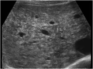
Transverse image of the liver with a diffuse inhomogeneous structure
Transverse image of the liver at a different level showing again the diffuse inhomogeneous structure
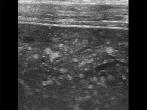
High resolution image of the same liver showing multiple hyperechoic lesions without any mass effect. The liver contour is normal
High resolution image of the same liver at a different level showing multiple hyperechoic lesions
Detail image f the same liver
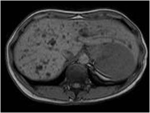
T1 MRI of the same liver. The lesions have a low signal intensity compared to the liver parechyma
T1 MRI of the same liver at another level. The lesions have a low signal intensity compared to the liver parechyma
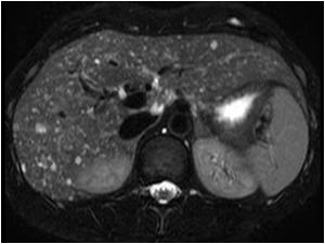
T2 MRI of the same liver. The lesions have a high signal intensity compared to the liver parechyma
T2 MRI of the same liver. at a different level. The lesions have a high signal intensity compared to the liver parechyma
cineloop

Conclusion

The intrahepatic lesions proved to be von Meyenburg complexes or biliary hamartomas
Biliary hamartomas or von Meyenburg complexes are uncommon benign malformations of the bile ducts. They are composed of disorganized bile ducts and sometimes fibrocollagenous stroma. They are usually multiple small (<1 cm.) round or irregular lesions scattered throughout the liver. These lesions are often discovered incidentally.

Imaging appearance often varies. On ultrasound, they may be hypoechoic, hyperechoic, or mixed echogenicity structures.  CT shows hypodense, small hepatic nodules, scattered throughout both liver lobes with no enhancement in most of the cases. On MRI, VMCs are numerous intrahepatic tiny cystoid lesions, reveal hypointensity on T1-weighted and hyperintensity on T2-weighted pulse sequences as compared to surrounding parenchyma

Details

  • Sex: Female
  • Age: 41

Created with

  • Fujifilm Ultrasound System