May 2011

Empty gallbladder and abnormal structures next to the gallbladder

Clinical information

Jaundice

Ultrasound Images & Clips

Abnormal structures around the gallbladder

Empty gallbladder and abnormal structures around the gallbladder
Longitudinal image of the empty gallbladder and abnormal structures.
Detail image of the structures next to the gallbladder showing tortuous veins
Color doppler image showing flow in the tortuous veins
Transverse image of left liver lobe showing dilatated intrahepatic bile ducts.
Another image of left liver lobe showing dilatated intrahepatic bile ducts.
The intrahepatic bile ducts of the right liver lobe are not so dilatated
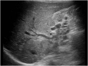
Detail of the mass with invasion en compression of the bile ducts and portal vein
Image of the liver hilum showing the obstruction of the portal vein and collateral veins in the gallbladder region
Detail image of the liver hilum showing tumor in the portal veins
Dtail image of the bile ducts with tumor masses
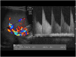
The hepatic artery is open and shows high peak systolic velocities of more than 2 mtrs. /sec. as can be expected in a case of portal vein occlusion
The pancreas of the patient was normal, but there were also collateral veins around the pancreas
Flow in the collateral veins around the pancreas
Axial CT arterial phase
Axial CT venous phase
Coronal CT venous phase
Sagittal CT venous phase

Conclusion

The ultrasound findings are a centrally located tumor mass in the liver with invasion and obstruction of the intrahepatic bile ducts of the left liver lobe. Portal vein thrombosis and collateral veins around the gallbladder and pancreas. Biopsy proved the mass to be a cholangiocarcinoma. A centrally in the liver located cholangiocarcinoma is called a Klatskin tumor

Details

  • Sex: Male
  • Age: 42

Created with

  • Fujifilm Ultrasound System