February 2012

Abnormal internal genitalia

Clinical information

Intermittent periodical pain in the lower abdomen. She has a normal menstrual cycle!
Ultrasound Images & Clips

Abnormal internal genitalia in a 14 year old girl

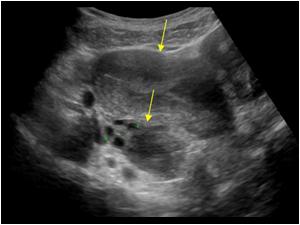
Transverse image of a cystic structure dorsal of the nearly empty bladder
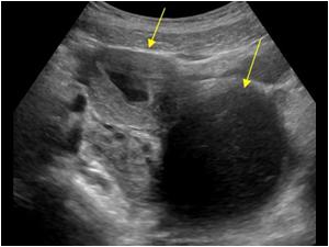
Longitudinal image of the lower abdomen and the cystic structure and a uterus with a fluid filled endometrial cavity
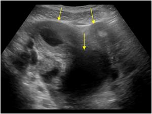
The solution of the normal menstrual cycle lies in this transverse image, where not only one but 2 uterine bodies are found
Another transverse omage showing two uterine bodies
Another transverse image
Longitudinal image of the normal uterine body and normal uterine cavity on the left side of the patient
Transverse image of the normal left uterine body and left ovary
A double uterus is called a uterus didelphys.This patient also had a double or septated vagina of which the right side was occluded causing the hematometra and hematocolpos
Transverse image of the normal left uterine body and left ovary A double uterus is called a uterus didelphys.This patient also had a double or septated vagina of which the right side was occluded causing the hematometra and hematocolpos
In cases of genital abnormalities always check the kidneys. No right kidney was found in this girl
The left kidney is normal but slightly larger than normal

Conclusion

The American Fertility Society (now American Society of Reproductive Medicine) Classification distinguishes:
Class I: Müllerian agenesis (absent uterus).
Uterus is not present, vagina only rudimentary or absent. The condition is also called Mayer-Rokitansky-Kuster-Hauser syndrome. The patient with MRKH syndrome will have primary amenorrhea.
Class II: Unicornuate uterus (a one-sided uterus).
Only one side of the Müllerian duct forms. The uterus has a typical "penis shape" on imaging systems.
Class III: Uterus didelphys, also uterus didelphis (double uterus).
Both Müllerian ducts develop but fail to fuse, thus the patient has a "double uterus". This may be a condition with a double cervix and a vaginal partition (v.i.), or the lower Müllerian system fused into its unpaired condition. See Triplet-birth with Uterus didelphys for a case of a woman having spontaneous birth in both wombs with twins.
Class IV: Bicornuate uterus (uterus with two horns).
Only the upper part of that part of the Müllerian system that forms the uterus fails to fuse, thus the caudal part of the uterus is normal, the cranial part is bifurcated. The uterus is "heart-shaped".
Class V: Septated uterus (uterine septum or partition).
The two Müllerian ducts have fused, but the partition between them is still present, splitting the system into two parts. With a complete septum the vagina, cervix and the uterus can be partitioned. Usually the septum affects only the cranial part of the uterus. A uterine septum is the most common uterine malformation and a cause for miscarriages.

The patient was referred to the gynaecologist who confirmed the diagnosis

Details

  • Sex: Female
  • Age: 14

Created with

  • Fujifilm Ultrasound System