July 2012

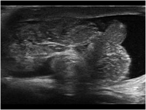
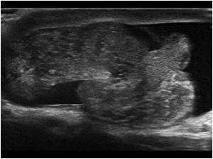
Vascularized right testis. Does this exclude a testicular torsion?

Clinical information

Acute onset of scrotal pain on the right side that started a few hours before the ultrasound examination. At the time of the examination the pain had already diminished a bit, but was still present
Ultrasound Images & Clips

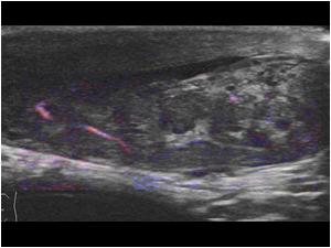
Vascularized normal looking testis. Does this exclude a testicular torsion?

Longitudinal image of the right testis with vessels seen with e-flow
Color doppler image of the left testis
Another color doppler image of the right testis. There are less vessels seen than on the left side
The right testis looks completely normal
The epididymis is slightly thickened and shows vessels with color doppler. Does this mean that there is an epididymitis?
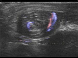
Transverse image of the epididymis and right testis with flow
A part of the epididymis has a spiral aspect. Does that change your diagnosis
Videoclip of the peritesticular structures
Color doppler image of the spiral stucture showing spiral flow
A spiral aspect always means that there is a torsion. This is an indication for immediate surgery. Sometimes there is some flow visible in the testis and or epididymis, when the tosion is not complete. However this can very easily change.
This is the epidydimis of a 13 year old boy with acute onset of scrotal pain. In this case there is not a spiral aspect but a knot
The testis is slightly swollen
Longitudinal image of the normal testis for comparison
In this case there is also minimal detactable flow
Is is possible to obtain a doppler signal but the velocities are extremely low
!8 year old with a painfull testis for a few hours
There is some flow detectable
The normal testis is not swollen and there are more vessels visible
The doppler signal has a low resistance signal
Again there is a knot visible in the transverse view indicating testicular torsion even though there is still flow detectable
Longitudinal image with the knot dorsal of the testis

Conclusion

All patients were operated and the diagnosis testicular torsion was confirmed. At the time of operation the testis in these patients was already discolored. The testis was detordated and recovered quickly in all cases. In cases of an incomplete testicular torsion the symptoms can change according to the amount of torsion. Flow can be detected and does not exclude a torsion. A spiral aspect of the pertesticular vessels or a peritesticular knot is always highly suspicious for a torsion. Also look for other signs like swelling and changes in the doppler signal

Details

  • Sex: Male
  • Age: 22

Created with

  • Fujifilm Ultrasound System