December 2013

Abnormal popliteal artery

Clinical information

Patient was referred to exclude a deep venous thrombosis. She had pain in the calf that progressed during walking. She could not walk without pain for more than 100 meters.
Ultrasound Images & Clips

Abnormal popliteal artery

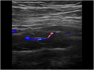
Longitudinal image of the popliteal artery showing a severe stenosis with a very narrow and irregular lumen
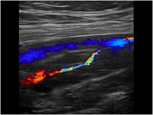
Another longitudinal image of the same popliteal artery with severe stenosis. The more superficial lying popliteal vein is patent
Transverse image of the same artery and vein
Detail image of the popliteal artery.
The stenosis is not caused by atherosclerotic changes
Detail image of the popliteal artery. The stenosis is not caused by atherosclerotic changes
transverse image of the popliteal artery showing cystic changes in the wall of the artery
MRA showing absent flow in the politeal artery
Detail of the popliteal area
MRI of the knee showing cystic lesions
MRI
MRI
MRI
Longitudinal image of the common femoral artery in an older male patient also showing cystic changes in the outer layer of the arterial wall. The intima layer is completely normal. The cystic changes cause minimal impression of the lumen
Longitudinal image of the common femoral artery showing the cystic changes
Transverse image of the common femoral artery showing the cystic changes
Longitudinal detail image of the cystic changes

Conclusion

The deep venous system in both patients was completely normal.
There was a significant stenosis in the popliteal artery of the first patient that was caused by cystic adventitial disease. The second older patient also had cystic adventitial changes but without a significant stenosis.

Details

  • Sex: Female
  • Age: 50

Created with

  • Fujifilm Ultrasound System