August 2018

Abnormal supraspinatus tendon

Clinical information

Middle age female patient with shoulder pain examined for SONOZORG a primary care ultrasound facility.

How would you describe it?
A) tendinosis
B) tendinosis and partial articular sided rupture
C) Tendinosis and intratendinous rupture
D) full thickness rupture
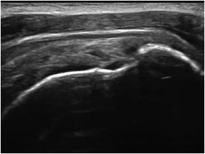
Ultrasound Images & Clips

Abnormal supraspinatus tendon.

a
a
Another longitudinal image
A third longitudinal image
A transverse image showing cortical irregularities
This is a longitudinal image of the supraspinatus tendon of another patient. It is not always possible to determine the exact extent of a tendon rupture. There is a partial rupture and volume loss and bursal thickening
Transverse image of the same patient
Always check if the patient has been operated!
This tendon also shows volume loss but the tendon rupture has been repaired and the bony anchors are visible
Always check if the patient has been operated! This tendon also shows volume loss but the tendon rupture has been repaired and the bony anchors are visible
Another longitudinal image of the operated patient
The tendon shows volume loss and the bursa is thickened. But the tendon is still intact

Conclusion

The first patient had some tendinosis and a partial articular sided supraspinatus tendon rupture. The rupture communicates with the articular cartilage but not with the bursa. There are also cortical irregularities
The second patient also had a supraspinatus tendon rupture with volume loss and bursal thickening but the exact extent was unclear (intratendinous or partial bursal sided).
The third patient also showed volume loss of the tendon but was operated on the tendon which was still intact.

Details

  • Sex: Female
  • Body part: Shoulder

Created with

  • Fujifilm Ultrasound System