Since 3 or 4 months this patient had bilateral knee pain, left more compared to right. The complaints started gradually and increased in time. Around the time of onset, the patient was renovating her house and did a lot of heavy lifting and working on her knees.
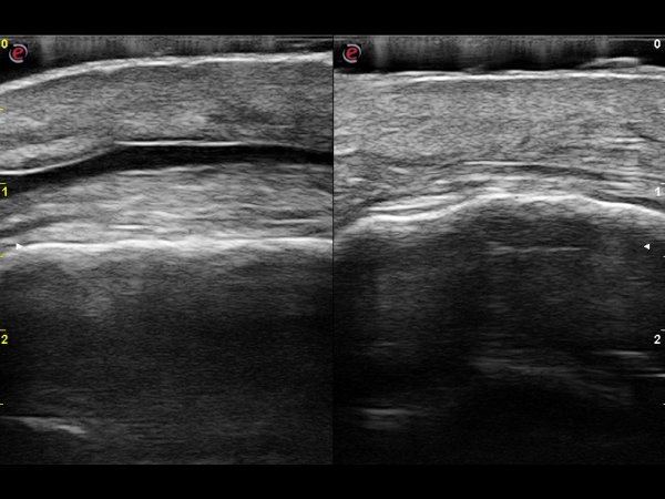
Pathology recognition or analysis was done with the help of the "SonoSkills pathology checklist". SHAPE: The prepatellar and superficial infrapatellar bursa have increased in size. This was only noted by using a gel stand-off to reduce the pressure of the transducer applied to the skin. ECHOGENICITY: an anechoic effusion fills both bursa's, most likely a fluid. A small anechoic zone can be seen superficial to the proximal patellar tendon, probably located in the distal part of the prepatellar bursa. CONTINUITY: there seem to be no signs of tearing a certain anatomical tissue. All fibers seem to be intact. DOPPLER: checking with power Doppler showed no signs of neovascularisation. FUNCTIONAL: The knee function was not impaired with ultrasound guided movements or functional loading. LAX/SAX: The findings could be seen in both longitudinal (LAX) and transverse (SAX) imaging. LEFT/RIGHT: ultrasound changes could only be seen in the left knee, not in the right knee.
")
")
")
")
I assessed this as a prepatellar and superficial infrapatellar bursitis of the left knee. The bursa's seem to be filled with fluid, and did not show signs of neovascularisation. The right knee did not show any ultrasound changes of the sonoanatomy. "The superficial infrapatellar bursa is localized in the soft tissue anterior to the distal third of the patellar tendon and overlying skin. Bursitis can be caused by chronic trauma due to occupational kneeling and direct trauma. Chronic hemorrhagic cases can calcify and simulate a soft tissue sarcoma. Viegas and others (2006) showed that this bursa can be located directly anterior to the tibial tuberosity but also can appear in a slightly superior position, or even be absent. The “normal” measurements of this bursa is respectively 19.5 mm, 21.2 mm and 2.2 mm. Communication between this bursa and the deep infrapatellar bursa around the lateral margin of the patellar tendon is sometimes possible".


Marc is founder of, and trainer at, SonoSkills. SonoSkills is an organization dedicated to MSK ultrasound education. He's also an MSK Sonographer at the Laurentius Hospital in Roermond, the Netherlands.
More Cases from Marc Schmitz

